


Crystal structure of calmodulin in complex with KN93 (1:1 complex)
Liu, X.C.To be published.
Experimental Data Snapshot
Starting Model: experimental
View more details
Entity ID: 1 | |||||
|---|---|---|---|---|---|
| Molecule | Chains | Sequence Length | Organism | Details | Image |
| Calmodulin-1 | 155 | Homo sapiens | Mutation(s): 0 Gene Names: CALM1, CALM, CAM, CAM1 | 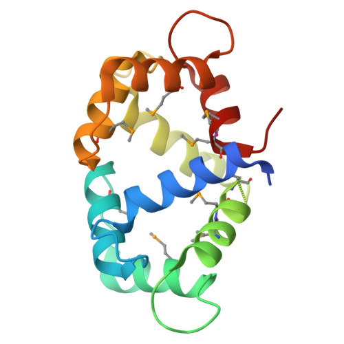 | |
UniProt & NIH Common Fund Data Resources | |||||
Find proteins for P0DP23 (Homo sapiens) Explore P0DP23 Go to UniProtKB: P0DP23 | |||||
PHAROS: P0DP23 GTEx: ENSG00000198668 | |||||
Entity Groups | |||||
| Sequence Clusters | 30% Identity50% Identity70% Identity90% Identity95% Identity100% Identity | ||||
| UniProt Group | P0DP23 | ||||
Sequence AnnotationsExpand | |||||
| |||||
| Ligands 2 Unique | |||||
|---|---|---|---|---|---|
| ID | Chains | Name / Formula / InChI Key | 2D Diagram | 3D Interactions | |
| KN9 (Subject of Investigation/LOI) Query on KN9 | H [auth A], M [auth B], R [auth C] | N-[2-[[[3-(4'-Chlorophenyl)-2-propenyl]methylamino]methyl]phenyl]-N-(2-hydroxyethyl)-4'-methoxybenzenesulfonamide C26 H29 Cl N2 O4 S LLLQTDSSHZREGW-AATRIKPKSA-N |  | ||
| CA Query on CA | D [auth A] E [auth A] F [auth A] G [auth A] I [auth B] | CALCIUM ION Ca BHPQYMZQTOCNFJ-UHFFFAOYSA-N |  | ||
| Modified Residues 1 Unique | |||||
|---|---|---|---|---|---|
| ID | Chains | Type | Formula | 2D Diagram | Parent |
| MSE Query on MSE | A, B, C | L-PEPTIDE LINKING | C5 H11 N O2 Se |  | MET |
| Length ( Å ) | Angle ( ˚ ) |
|---|---|
| a = 69.336 | α = 90 |
| b = 40.264 | β = 92.47 |
| c = 173.413 | γ = 90 |
| Software Name | Purpose |
|---|---|
| PHENIX | refinement |
| autoPX | data reduction |
| Aimless | data scaling |
| PHASER | phasing |
| Funding Organization | Location | Grant Number |
|---|---|---|
| National Science Foundation (NSF, China) | China | -- |


