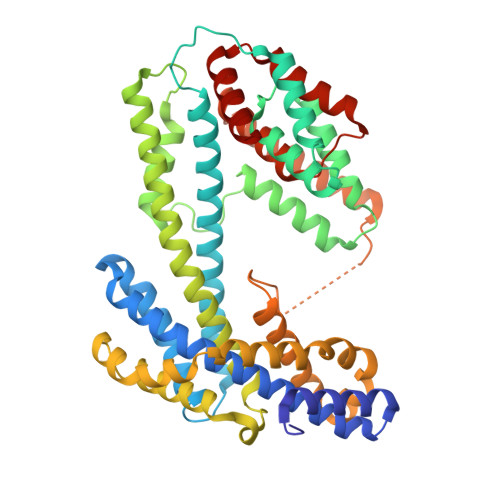
32UY | pdb_000032uy

Protein with 8.3 knot


Experimental Data Snapshot

  • Method: X-RAY DIFFRACTION
  • Resolution: 2.54 Å
  • R-Value Free: 
    0.255 (Depositor), 0.257 (DCC) 
  • R-Value Work: 
    0.219 (Depositor), 0.220 (DCC) 
  • R-Value Observed: 
    0.221 (Depositor) 

Starting Model: in silico
View more details

wwPDB Validation 3D Report Full Report

Validation slider image for 32UY

This is version 1.1 of the entry. See complete history. 

Literature

Unique features of EIGHT fold-Proteins with the most complex 8 3 knotted topology.

Mozajew, M., Sikora, M., Fortunka, M., Perlinska, A.P., Kluza, A., Niewieczerzal, S., da Silva, F.B., Lukaszewicz, M., Sikorska, J.A., Mikulska, L.J., Jackson, S.E., Sulkowska, J.I.

(2026) Proc Natl Acad Sci U S A 123: e2523017123-e2523017123

  • DOI: https://doi.org/10.1073/pnas.2523017123
  • Primary Citation Related Structures: 
    32UY, 9RH0, 9TMH

  • PubMed Abstract: 

    Herein, we present three crystal structures of the most complex knotted protein to date, an [Formula: see text] knot with eight crossings, which belongs to a clasp knot family. The fold of this protein is also unique, and we call it the EIGHT fold (Extraordinarily Intricate Globular Helical Tangled fold). The [Formula: see text] knot is found only in a certain group of bacteria, where it is strictly conserved, despite sequence identities as low as 10%. The [Formula: see text] structure shows a high level of internal symmetry, with structurally analogous halves. It is possible that the EIGHT fold evolved from a single unknotted "half" that duplicated and formed a complex protein. Based on an in vitro study of three selected [Formula: see text] proteins, we estimated their thermal and thermodynamic stability, and showed that they follow a two-state unfolding model under equilibrium conditions and their refolding occurs to a compact, nonnative state. Unfolding kinetics of [Formula: see text] knotted proteins reveal a complex, multiphase process spanning milliseconds to days. Six kinetic phases were resolved using stopped-flow fluorescence and far-ultraviolet absorbance circular dichroism spectroscopy, highlighting both rapid early events and slower rearrangements in secondary and tertiary structures. Finally, with a structure-based model, we showed that proteins with an [Formula: see text] knot can self-tie through a [Formula: see text] knotted intermediate state. They can also self-untie, albeit via an additional intermediate knot such as [Formula: see text]. Nevertheless, these proteins do not seem to fit with current folding theories known to date and require the development of a new approach.


  • Organizational Affiliation: 
    • Centre of New Technologies, University of Warsaw, Banacha 2C St., Warszawa 02-097, Poland.

Macromolecule Content 

  • Total Structure Weight: 108.89 kDa 
  • Atom Count: 7,059 
  • Modeled Residue Count: 906 
  • Deposited Residue Count: 944 
  • Unique protein chains: 1

Macromolecules

Find similar proteins by:|  3D Structure
Entity ID: 1
MoleculeChains  Sequence LengthOrganismDetailsImage
Protein with 8.3 knot
A, B
472Lachnospiraceae bacteriumMutation(s): 0 
Entity Groups
Sequence Clusters30% Identity50% Identity70% Identity90% Identity95% Identity100% Identity
Sequence Annotations
Expand
Reference Sequence

Small Molecules

Ligands 3 Unique
IDChains Name / Formula / InChI Key2D Diagram3D Interactions
PEG

Query on PEG


Download:Ideal Coordinates CCD File
I [auth B]DI(HYDROXYETHYL)ETHER
C4 H10 O3
MTHSVFCYNBDYFN-UHFFFAOYSA-N
SO4

Query on SO4


Download:Ideal Coordinates CCD File
E [auth A]
F [auth A]
G [auth A]
H [auth A]
J [auth B]
E [auth A],
F [auth A],
G [auth A],
H [auth A],
J [auth B],
K [auth B]
SULFATE ION
O4 S
QAOWNCQODCNURD-UHFFFAOYSA-L
EDO

Query on EDO


Download:Ideal Coordinates CCD File
C [auth A],
D [auth A]
1,2-ETHANEDIOL
C2 H6 O2
LYCAIKOWRPUZTN-UHFFFAOYSA-N

Experimental Data & Validation

Experimental Data

  • Method: X-RAY DIFFRACTION
  • Resolution: 2.54 Å
  • R-Value Free:  0.255 (Depositor), 0.257 (DCC) 
  • R-Value Work:  0.219 (Depositor), 0.220 (DCC) 
  • R-Value Observed: 0.221 (Depositor) 
Space Group: P 1 21 1
Unit Cell:
Length ( Å )Angle ( ˚ )
a = 69.38α = 90
b = 92.54β = 91.06
c = 118.54γ = 90
Software Package:
Software NamePurpose
PHENIXrefinement
MxCuBEdata collection
XDSdata reduction
XDSdata scaling
PHASERphasing
PHENIXmodel building
Cootmodel building

Structure Validation

View Full Validation Report


Entry History 

& Funding Information

Deposition Data


Funding OrganizationLocationGrant Number
Polish National Science CentrePoland2021/43/I/NZ1/03341

Revision History  (Full details and data files)

  • Version 1.0: 2026-10-21
    Type: Initial release
  • Version 1.1: 2026-10-28
    Changes: Database references