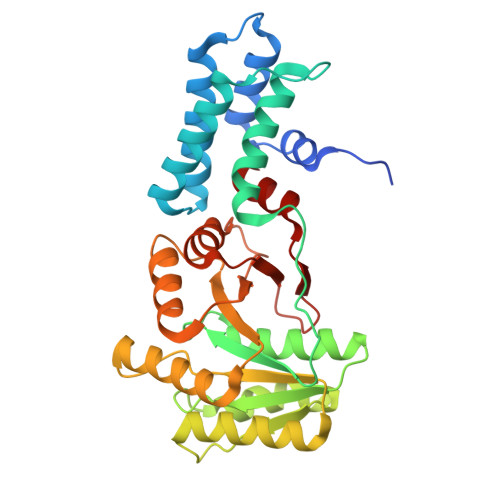
9YY5 | pdb_00009yy5

Crystal structure of P. furiosus SRP54 NG domain G225E


Experimental Data Snapshot

  • Method: X-RAY DIFFRACTION
  • Resolution: 1.70 Å
  • R-Value Free: 
    0.217 (Depositor), 0.217 (DCC) 
  • R-Value Work: 
    0.210 (Depositor), 0.210 (DCC) 
  • R-Value Observed: 
    0.210 (Depositor) 

Starting Model: experimental
View more details

wwPDB Validation 3D Report Full Report

Validation slider image for 9YY5

Ligand Structure Quality Assessment 


This is version 1.1 of the entry. See complete history. 

Literature

Structure and Function of Archaeal SRP54 Protein Mutants Associated with Human Severe Congenital Neutropenia.

Yang, M., Brim, A.D., Long, L.F., Wu, G.C., Schlichting, G.M., Denard, C.A., Bruner, S.D.

(2026) ACS Omega 11: 63170-63178

  • DOI: https://doi.org/10.1021/acsomega.6c06315
  • Primary Citation Related Structures: 
    9YDH, 9YY5, 9ZG6, 9ZJM

  • PubMed Abstract: 

    SRP54, a key component of the signal recognition particle, is a highly conserved enzyme both structurally and functionally. The protein plays a vital role in cellular protein translocation by interacting with the translating ribosome and a membrane receptor, thereby aiding protein folding and trafficking. Recent studies have shown that mutations in the SRP54 gene cause severe neutropenia and are associated with an increased risk of pediatric leukemias. Here, we present high-resolution structures of clinically relevant SRP54 mutant proteins from the archaea Pyrococcus furiosus . This work complements existing structural and functional data on SRP54 and offers a high-resolution structural basis for clinically relevant congenital mutations. The structures of three mutants are presented along with the model wild type. Overall, the results provide a detailed understanding of how mutations affect SRP54 structure. Additionally, comparing our findings with available structures from higher eukaryotes offers new insights into the mechanisms of dysfunction.


  • Organizational Affiliation: 
    • University of Florida, Department of Chemistry, Gainesville, Florida 32611, United States.

Macromolecule Content 

  • Total Structure Weight: 35.37 kDa 
  • Atom Count: 2,621 
  • Modeled Residue Count: 303 
  • Deposited Residue Count: 316 
  • Unique protein chains: 1

Macromolecules

Find similar proteins by:|  3D Structure
Entity ID: 1
MoleculeChains  Sequence LengthOrganismDetailsImage
Signal recognition particle 54 kDa protein316Pyrococcus furiosusMutation(s): 1 
Gene Names: srp54, PF1731
EC: 3.6.5.4
UniProt
Find proteins for Q8U070 (Pyrococcus furiosus (strain ATCC 43587 / DSM 3638 / JCM 8422 / Vc1))
Explore Q8U070 
Go to UniProtKB:  Q8U070
Entity Groups
Sequence Clusters30% Identity50% Identity70% Identity90% Identity95% Identity100% Identity
UniProt GroupQ8U070
Sequence Annotations
Expand
Reference Sequence

Experimental Data & Validation

Experimental Data

  • Method: X-RAY DIFFRACTION
  • Resolution: 1.70 Å
  • R-Value Free:  0.217 (Depositor), 0.217 (DCC) 
  • R-Value Work:  0.210 (Depositor), 0.210 (DCC) 
  • R-Value Observed: 0.210 (Depositor) 
Space Group: P 43 21 2
Unit Cell:
Length ( Å )Angle ( ˚ )
a = 73.249α = 90
b = 73.249β = 90
c = 123.689γ = 90
Software Package:
Software NamePurpose
PHENIXrefinement
Aimlessdata scaling
XDSdata reduction
PHENIXphasing
Cootmodel building

Structure Validation

View Full Validation Report


Ligand Structure Quality Assessment 


Entry History 

& Funding Information

Deposition Data


Funding OrganizationLocationGrant Number
Other governmentUnited States23L04

Revision History  (Full details and data files)

  • Version 1.0: 2026-10-28
    Type: Initial release
  • Version 1.1: 2026-11-04
    Changes: Database references