 Download Mendeley
Crystal structure of E19A mutant chitosanase from Bacillus subtilis MY002 complexed with GlcNs.
Xie, J., Xie, T., Wang, G.G.To be published.
Experimental Data Snapshot
Starting Model: experimental
View more details
wwPDB Validation 3D Report Full Report

Entity ID: 1 | |||||
|---|---|---|---|---|---|
| Molecule | Chains | Sequence Length | Organism | Details | Image |
| Chitosanase | 242 | Bacillus subtilis subsp. subtilis str. 168 | Mutation(s): 1 Gene Names: csn, BSU26890 EC: 3.2.1.132 | 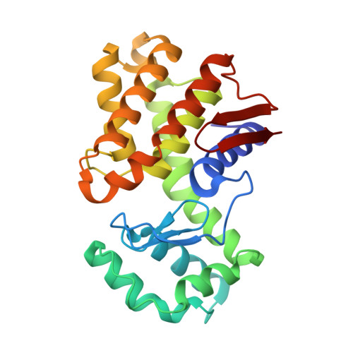 | |
UniProt | |||||
Entity Groups | |||||
| Sequence Clusters | 30% Identity50% Identity70% Identity90% Identity95% Identity100% Identity | ||||
| UniProt Group | O07921 | ||||
Sequence AnnotationsExpand | |||||
Reference Sequence | |||||
Entity ID: 2 | |||||
|---|---|---|---|---|---|
| Molecule | Chains | Length | 2D Diagram | Glycosylation | D Interactions |
| 2-amino-2-deoxy-beta-D-glucopyranose-(1-4)-2-amino-2-deoxy-beta-D-glucopyranose-(1-4)-2-amino-2-deoxy-beta-D-glucopyranose-(1-4)-(2~{R},3~{S},4~{R},5~{R},6~{R})-5-azanyl-6-(dioxidanyl)-2-(hydroxymethyl)oxane-3,4-diol | B | 4 | N/A | N/A | |
Entity ID: 3 | |||||
|---|---|---|---|---|---|
| Molecule | Chains | Length | 2D Diagram | Glycosylation | D Interactions |
| 2-amino-2-deoxy-beta-D-glucopyranose-(1-4)-2-amino-2-deoxy-beta-D-glucopyranose-(1-4)-2-amino-2-deoxy-beta-D-glucopyranose-(1-4)-2-amino-2-deoxy-beta-D-glucopyranose | C | 4 | N/A | N/A | |
| Length ( Å ) | Angle ( ˚ ) |
|---|---|
| a = 46.671 | α = 90 |
| b = 77.047 | β = 90 |
| c = 79.278 | γ = 90 |
| Software Name | Purpose |
|---|---|
| PHENIX | refinement |
| HKL-3000 | data scaling |
| HKL-3000 | data reduction |
| PHASER | phasing |
| PDB_EXTRACT | data extraction |
| Funding Organization | Location | Grant Number |
|---|---|---|
| Not funded | -- |
