







 Download Mendeley
Crystal structure of NUDIX hydrolase E83A mutant from Limisphaera ngatamarikiensis in complex with ITP
Wu, W.Q., Li, Z.X.To be published.
Experimental Data Snapshot
Starting Model: in silico
View more details
Entity ID: 1 | |||||
|---|---|---|---|---|---|
| Molecule | Chains | Sequence Length | Organism | Details | Image |
| NUDIX domain-containing protein | 211 | Limisphaera ngatamarikiensis | Mutation(s): 1 Gene Names: G4L39_03320 | 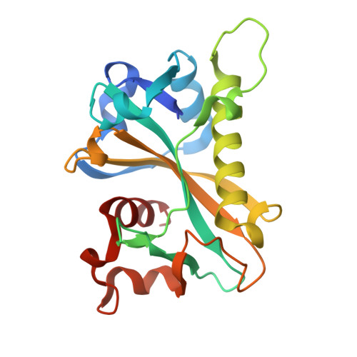 | |
UniProt | |||||
Find proteins for A0A6M1RLF6 (Limisphaera ngatamarikiensis) Explore A0A6M1RLF6 Go to UniProtKB: A0A6M1RLF6 | |||||
Entity Groups | |||||
| Sequence Clusters | 30% Identity50% Identity70% Identity90% Identity95% Identity100% Identity | ||||
| UniProt Group | A0A6M1RLF6 | ||||
Sequence AnnotationsExpand | |||||
Reference Sequence | |||||
| Ligands 2 Unique | |||||
|---|---|---|---|---|---|
| ID | Chains | Name / Formula / InChI Key | 2D Diagram | 3D Interactions | |
| CZU (Subject of Investigation/LOI) Download:Ideal Coordinates CCD File | E [auth A], G [auth B], I [auth C], K [auth D] | [[(2~{R},3~{S},4~{R},5~{R})-3,4-bis(oxidanyl)-5-(6-oxidanylidene-1~{H}-purin-9-yl)oxolan-2-yl]methoxy-oxidanyl-phosphoryl] phosphono hydrogen phosphate C10 H15 N4 O14 P3 HAEJPQIATWHALX-KQYNXXCUSA-N | 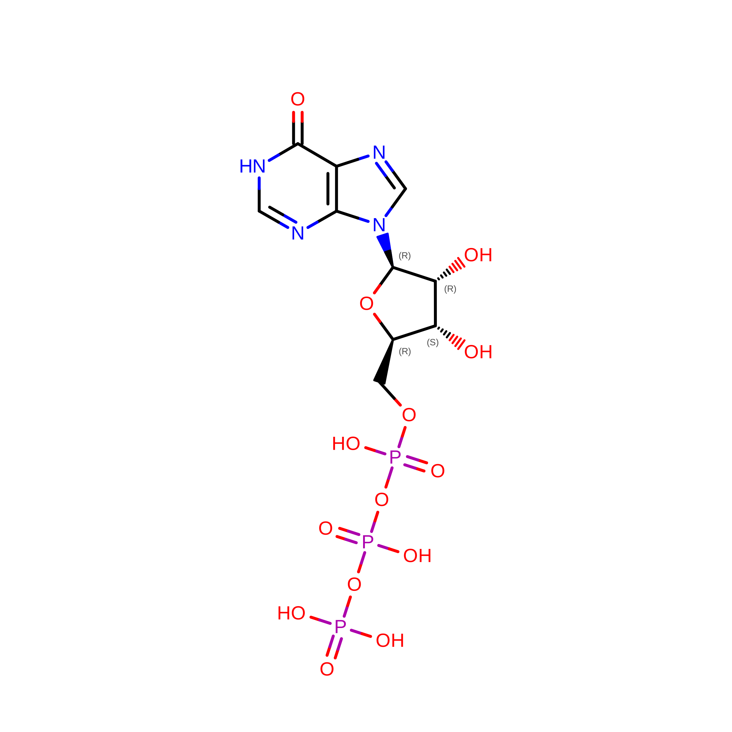 | ||
| ZN (Subject of Investigation/LOI) Download:Ideal Coordinates CCD File | F [auth A], H [auth B], J [auth C], L [auth D] | ZINC ION Zn PTFCDOFLOPIGGS-UHFFFAOYSA-N |  | ||
| Length ( Å ) | Angle ( ˚ ) |
|---|---|
| a = 148.317 | α = 90 |
| b = 70.695 | β = 90 |
| c = 81.755 | γ = 90 |
| Software Name | Purpose |
|---|---|
| PHENIX | refinement |
| PDB_EXTRACT | data extraction |
| XDS | data scaling |
| PHENIX | phasing |
| Aimless | data scaling |
| XDS | data reduction |
| Funding Organization | Location | Grant Number |
|---|---|---|
| National Natural Science Foundation of China (NSFC) | China | 82304614 |








