


Design and synthesis of 7-aryl-6-fluoro-2-aminoquinolines as potent and highly selective human neuronal nitric oxide synthase inhibitors
Ansari, A., Rathnayake, A.D., Li, H., Hardy, C.D., Poulos, T.L., Silverman, R.B.To be published.
Experimental Data Snapshot
Starting Model: experimental
View more details
Entity ID: 1 | |||||
|---|---|---|---|---|---|
| Molecule | Chains | Sequence Length | Organism | Details | Image |
| Nitric oxide synthase, brain | 423 | Homo sapiens | Mutation(s): 2 Gene Names: NOS1 EC: 1.14.13.39 | 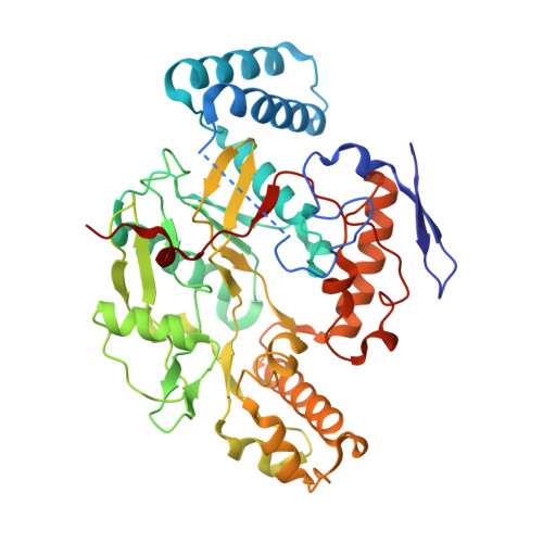 | |
UniProt & NIH Common Fund Data Resources | |||||
Find proteins for P29475 (Homo sapiens) Explore P29475 Go to UniProtKB: P29475 | |||||
PHAROS: P29475 GTEx: ENSG00000089250 | |||||
Entity Groups | |||||
| Sequence Clusters | 30% Identity50% Identity70% Identity90% Identity95% Identity100% Identity | ||||
| UniProt Group | P29475 | ||||
Sequence AnnotationsExpand | |||||
| |||||
| Ligands 5 Unique | |||||
|---|---|---|---|---|---|
| ID | Chains | Name / Formula / InChI Key | 2D Diagram | 3D Interactions | |
| HEM Query on HEM | E [auth A], K [auth B], O [auth C], U [auth D] | PROTOPORPHYRIN IX CONTAINING FE C34 H32 Fe N4 O4 KABFMIBPWCXCRK-RGGAHWMASA-L |  | ||
| A1BUF (Subject of Investigation/LOI) Query on A1BUF | G [auth A], M [auth B], Q [auth C], W [auth D] | (2M)-2-(2-amino-6-fluoro-4-methylquinolin-7-yl)-5-(aminomethyl)phenol C17 H16 F N3 O HSYNPKNGWVSVIB-UHFFFAOYSA-N |  | ||
| H4B Query on H4B | F [auth A], L [auth B], P [auth C], V [auth D] | 5,6,7,8-TETRAHYDROBIOPTERIN C9 H15 N5 O3 FNKQXYHWGSIFBK-RPDRRWSUSA-N |  | ||
| GOL Query on GOL | H [auth A] I [auth A] N [auth B] R [auth C] S [auth C] | GLYCEROL C3 H8 O3 PEDCQBHIVMGVHV-UHFFFAOYSA-N |  | ||
| ZN Query on ZN | J [auth B], T [auth C] | ZINC ION Zn PTFCDOFLOPIGGS-UHFFFAOYSA-N |  | ||
| Length ( Å ) | Angle ( ˚ ) |
|---|---|
| a = 118.881 | α = 90 |
| b = 52.226 | β = 90 |
| c = 164.574 | γ = 90 |
| Software Name | Purpose |
|---|---|
| PHENIX | refinement |
| Aimless | data scaling |
| XDS | data reduction |
| REFMAC | phasing |
| Funding Organization | Location | Grant Number |
|---|---|---|
| National Institutes of Health/National Institute of General Medical Sciences (NIH/NIGMS) | United States | GM131920 |


