


Structural Analysis of Inhibitor Binding to the Feline Enteric Coronavirus (FECV) Main Protease
Maryam, A., Moquin, S.A., Dovala, D., Kaur, J., Kurt Yilmaz, N., Shaqra, A.M., Schiffer, C.A.(2025) Viruses 17
Experimental Data Snapshot
Starting Model: experimental
View more details
(2025) Viruses 17
Entity ID: 1 | |||||
|---|---|---|---|---|---|
| Molecule | Chains | Sequence Length | Organism | Details | Image |
| Peptidase C30 | 302 | Feline coronavirus UU23 | Mutation(s): 0 | 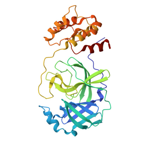 | |
UniProt | |||||
Find proteins for D3KDL2 (Feline coronavirus UU23) Explore D3KDL2 Go to UniProtKB: D3KDL2 | |||||
Entity Groups | |||||
| Sequence Clusters | 30% Identity50% Identity70% Identity90% Identity95% Identity100% Identity | ||||
| UniProt Group | D3KDL2 | ||||
Sequence AnnotationsExpand | |||||
| |||||
| Ligands 2 Unique | |||||
|---|---|---|---|---|---|
| ID | Chains | Name / Formula / InChI Key | 2D Diagram | 3D Interactions | |
| 4WI (Subject of Investigation/LOI) Query on 4WI | E [auth A], F [auth B], I [auth C], J [auth D] | (1R,2S,5S)-N-{(1E,2S)-1-imino-3-[(3S)-2-oxopyrrolidin-3-yl]propan-2-yl}-6,6-dimethyl-3-[3-methyl-N-(trifluoroacetyl)-L-valyl]-3-azabicyclo[3.1.0]hexane-2-carboxamide C23 H34 F3 N5 O4 WDVIRQQKRMIXGS-XIFHJVQQSA-N |  | ||
| PEG Query on PEG | G [auth B], H [auth B] | DI(HYDROXYETHYL)ETHER C4 H10 O3 MTHSVFCYNBDYFN-UHFFFAOYSA-N |  | ||
| Length ( Å ) | Angle ( ˚ ) |
|---|---|
| a = 63.919 | α = 90 |
| b = 106.316 | β = 90.51 |
| c = 78.509 | γ = 90 |
| Software Name | Purpose |
|---|---|
| PHENIX | refinement |
| Coot | model building |
| XDS | data reduction |
| XDS | data scaling |
| PHASER | phasing |
| Funding Organization | Location | Grant Number |
|---|---|---|
| Other private | -- |


