9LMC | pdb_00009lmc

Crystal structure of Guanine-II riboswitch in complex with guanine soaked with Mn2+


Experimental Data Snapshot

  • Method: X-RAY DIFFRACTION
  • Resolution: 2.97 Å
  • R-Value Free: 
    0.273 (Depositor), 0.269 (DCC) 
  • R-Value Work: 
    0.226 (Depositor), 0.226 (DCC) 
  • R-Value Observed: 
    0.231 (Depositor) 

Starting Model: experimental
View more details

wwPDB Validation   3D Report Full Report


Ligand Structure Quality Assessment 


This is version 1.0 of the entry. See complete history


Literature

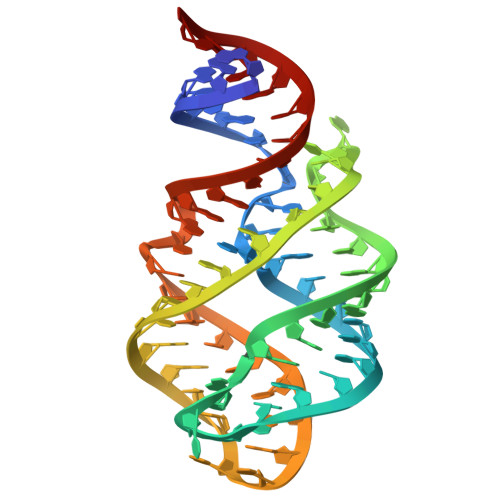
Ligand specificity and adaptability revealed by the first Guanine-II riboswitch tertiary structure.

Li, H.Shen, X.Xu, X.Tai, X.He, M.Zhang, J.Ren, A.

(2025) Nucleic Acids Res 53

  • DOI: https://doi.org/10.1093/nar/gkaf884
  • Primary Citation of Related Structures:  
    9LJN, 9LKC, 9LKE, 9LKF, 9LMC, 9V4U, 9V4V, 9V4X, 9V4Y, 9V4Z, 9V50, 9V51

  • PubMed Abstract: 

    A comprehensive understanding of the fundamental principles governing RNA-small molecule interactions is crucial for advancing RNA-targeting therapeutics with small molecules. Riboswitches, a class of noncoding RNAs, regulate gene expression by direct interaction with small-molecule metabolites. In this work, we report an in-depth structure-based investigation of a newly identified riboswitch, Guanine-II, which, despite sharing a conserved scaffold with the Guanine-I riboswitch, exhibits strikingly distinct small molecule ligand-binding characteristics. Through a comprehensive structural analysis of the Guanine-II riboswitch bound to various guanine analogs, combined with comparative studies of other guanine riboswitch variants, including Guanine-I and Xanthine-II riboswitches, as well as isothermal titration calorimetry, we reveal local structural rearrangements that precisely modulate small-molecule ligand adaptability. We further demonstrate that subtle differences in the composition and peripheral architecture of the binding pocket are key determinants of ligand-binding specificity. Additionally, based on the similarity in ligand recognition patterns with the tetrahydrofolate-II riboswitch, we identified additional compounds that bind to the Guanine-II riboswitch through a structure-guided rational search, providing valuable structural insights for the discovery of small molecules targeting RNA.


  • Organizational Affiliation
    • Department of Cardiology of The Second Affiliated Hospital and Life Sciences Institute and School of Medicine, Zhejiang University, Hangzhou 310058, China.

Macromolecules
Find similar nucleic acids by:  (by identity cutoff)  |  3D Structure
Entity ID: 1
MoleculeChains LengthOrganismImage
PRTg (71-MER)
A, B
71Paenibacillus sp. FSL E2-0178
Sequence Annotations
Expand
  • Reference Sequence
Small Molecules
Ligands 4 Unique
IDChains Name / Formula / InChI Key2D Diagram3D Interactions
GTP
Query on GTP

Download Ideal Coordinates CCD File 
K [auth B]GUANOSINE-5'-TRIPHOSPHATE
C10 H16 N5 O14 P3
XKMLYUALXHKNFT-UUOKFMHZSA-N
GUN (Subject of Investigation/LOI)
Query on GUN

Download Ideal Coordinates CCD File 
C [auth A],
J [auth B]
GUANINE
C5 H5 N5 O
UYTPUPDQBNUYGX-UHFFFAOYSA-N
MN
Query on MN

Download Ideal Coordinates CCD File 
D [auth A]
E [auth A]
F [auth A]
G [auth A]
H [auth A]
D [auth A],
E [auth A],
F [auth A],
G [auth A],
H [auth A],
I [auth A],
L [auth B],
M [auth B],
N [auth B]
MANGANESE (II) ION
Mn
WAEMQWOKJMHJLA-UHFFFAOYSA-N
MG
Query on MG

Download Ideal Coordinates CCD File 
O [auth B]MAGNESIUM ION
Mg
JLVVSXFLKOJNIY-UHFFFAOYSA-N
Experimental Data & Validation

Experimental Data

  • Method: X-RAY DIFFRACTION
  • Resolution: 2.97 Å
  • R-Value Free:  0.273 (Depositor), 0.269 (DCC) 
  • R-Value Work:  0.226 (Depositor), 0.226 (DCC) 
  • R-Value Observed: 0.231 (Depositor) 
Space Group: P 2 2 21
Unit Cell:
Length ( Å )Angle ( ˚ )
a = 119.425α = 90
b = 37.719β = 90
c = 94.496γ = 90
Software Package:
Software NamePurpose
PHENIXrefinement
XDSdata scaling
autoPROCdata reduction
PHENIXphasing

Structure Validation

View Full Validation Report


Ligand Structure Quality Assessment 


Entry History & Funding Information

Deposition Data


Funding OrganizationLocationGrant Number
National Natural Science Foundation of China (NSFC)China--

Revision History  (Full details and data files)

  • Version 1.0: 2025-11-26
    Type: Initial release