9IQQ | pdb_00009iqq

Crystal structure of PKM2 in complex with a natural activator


Experimental Data Snapshot

  • Method: X-RAY DIFFRACTION
  • Resolution: 2.70 Å
  • R-Value Free: 
    0.264 (Depositor), 0.266 (DCC) 
  • R-Value Work: 
    0.256 (Depositor), 0.257 (DCC) 

Starting Model: experimental
View more details

wwPDB Validation   3D Report Full Report


Ligand Structure Quality Assessment 


This is version 1.0 of the entry. See complete history


Literature

GSH as an A-A Type Allosteric Activator of PKM2: Modulating Cancer Cell Homeostasis and Ferroptosis Susceptibility.

Chen, T.J.Lo, C.J.Wu, M.J.Sit, W.Y.Hsu, H.Y.Huang, Y.C.Lu, C.H.Chen, Y.L.Fang, W.K.Yang, S.M.Chen, P.L.Haraguchi, T.Hiraoka, Y.Lin, C.Y.Cheng, M.L.Yang, M.H.Kung, H.J.Wang, W.C.

(2025) Adv Sci (Weinh) : e19368-e19368

  • DOI: https://doi.org/10.1002/advs.202519368
  • Primary Citation of Related Structures:  
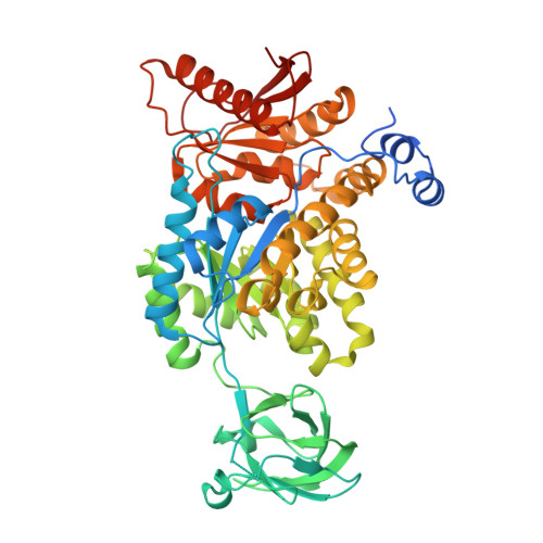
    9IQQ

  • PubMed Abstract: 

    This study identifies glutathione (GSH) as an endogenous A-A type allosteric activator of pyruvate kinase M2 (PKM2), stabilizing it in its active tetrameric form through binding at the A-A interface. This PKM2-GSH interaction links GSH metabolism to ferroptosis regulation. Transcriptomic analyses across cancers demonstrate strong correlations between GSH, SLC7A11, PKM2, glycolysis, and ferroptosis pathways. By depleting GSH and activating PKM2, ferroptosis is enhanced in PKM2-dependent cancer models. This approach leads to significant changes in central carbon and lipid metabolism, disrupts mitochondrial function, and drives ferroptotic cell death. The combined treatment markedly suppresses tumor growth in a xenograft model. Elevated PKM2 and SLC7A11 expression levels correlate with poorer survival outcomes, indicating their potential as biomarkers for ferroptosis-based therapy. The findings demonstrate a dual role for GSH in cellular homeostasis and identify the PKM2-GSH-SLC7A11 axis as a therapeutic target for aggressive cancers.


  • Organizational Affiliation
    • Institute of Molecular and Cellular Biology and Department of Life Science, National Tsing Hua University, Hsinchu, 300044, Taiwan.

Macromolecules
Find similar proteins by:  (by identity cutoff)  |  3D Structure
Entity ID: 1
MoleculeChains Sequence LengthOrganismDetailsImage
Pyruvate kinase PKM
A, B, C, D
551Homo sapiensMutation(s): 0 
Gene Names: PKMOIP3PK2PK3PKM2
EC: 2.7.1.40 (PDB Primary Data), 2.7.11.1 (PDB Primary Data), 2.7.10.2 (PDB Primary Data)
UniProt & NIH Common Fund Data Resources
Find proteins for P14618 (Homo sapiens)
Explore P14618 
Go to UniProtKB:  P14618
PHAROS:  P14618
GTEx:  ENSG00000067225 
Entity Groups  
Sequence Clusters30% Identity50% Identity70% Identity90% Identity95% Identity100% Identity
UniProt GroupP14618
Sequence Annotations
Expand
  • Reference Sequence
Experimental Data & Validation

Experimental Data

  • Method: X-RAY DIFFRACTION
  • Resolution: 2.70 Å
  • R-Value Free:  0.264 (Depositor), 0.266 (DCC) 
  • R-Value Work:  0.256 (Depositor), 0.257 (DCC) 
Space Group: P 1 21 1
Unit Cell:
Length ( Å )Angle ( ˚ )
a = 73.824α = 90
b = 130.288β = 93.969
c = 104.456γ = 90
Software Package:
Software NamePurpose
REFMACrefinement
HKL-2000data reduction
HKL-2000data scaling
MOLREPphasing

Structure Validation

View Full Validation Report


Ligand Structure Quality Assessment 


Entry History & Funding Information

Deposition Data


Funding OrganizationLocationGrant Number
National Science Council (NSC, Taiwan)TaiwanNSC-105-2314-B-007 -001

Revision History  (Full details and data files)

  • Version 1.0: 2025-11-19
    Type: Initial release