36OU | pdb_000036ou

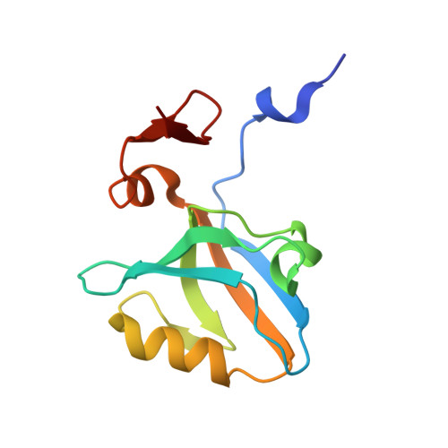
Room-temperature multiconformer model of PSD-95 PDZ3-CRIPT


Experimental Data Snapshot

  • Method: X-RAY DIFFRACTION
  • Resolution: 1.35 Å
  • R-Value Free: 
    0.147 (Depositor), 0.149 (DCC) 
  • R-Value Work: 
    0.122 (Depositor), 0.122 (DCC) 
  • R-Value Observed: 
    0.124 (Depositor) 

Starting Model: experimental
View more details

wwPDB Validation 3D Report Full Report

Validation slider image for 36OU

This is version 1.0 of the entry. See complete history. 

Literature

Symmetry Breaking under Vectorial Perturbations in Macromolecular X-ray Crystallography.

Wang, H.K., Greisman, J.B., Dalton, K.M., Hewitt, R.A., White, K.I., Hekstra, D.R.

(2026) bioRxiv 

  • DOI: https://doi.org/10.64898/2026.09.28.753211
  • Primary Citation Related Structures: 
    36OR, 36OU

  • PubMed Abstract: 

    Proteins are molecular machines whose dynamics govern their function. Using time-resolved crystallography, the basis of protein mechanics can be uncovered by physical perturbation on protein crystals. Perturbations are often vector fields that, in crystals, break symmetry. Lacking existing tools for analyzing symmetry breaking, we built open-source software to do so. We applied this method to three time-resolved crystallography experiments and analyzed the effect of vectorial perturbations on macromolecular crystal structure. We demonstrate that our method can handle symmetry reduction in all 65 Sohncke space groups.


  • Organizational Affiliation: 
    • Department of Molecular and Cellular Biology, Harvard University, Cambridge, MA 02138, USA.

Macromolecule Content 

  • Total Structure Weight: 13.81 kDa 
  • Atom Count: 1,616 
  • Modeled Residue Count: 126 
  • Deposited Residue Count: 128 
  • Unique protein chains: 2

Macromolecules

Find similar proteins by:|  3D Structure
Entity ID: 1
MoleculeChains  Sequence LengthOrganismDetailsImage
Disks large homolog 4119Rattus norvegicusMutation(s): 0 
Gene Names: Dlg4, Dlgh4, Psd95
UniProt
Find proteins for P31016 (Rattus norvegicus)
Explore P31016 
Go to UniProtKB:  P31016
Entity Groups
Sequence Clusters30% Identity50% Identity70% Identity90% Identity95% Identity100% Identity
UniProt GroupP31016
Sequence Annotations
Expand
Reference Sequence
Find similar proteins by:  Sequence   |   3D Structure  
Entity ID: 2
MoleculeChains  Sequence LengthOrganismDetailsImage
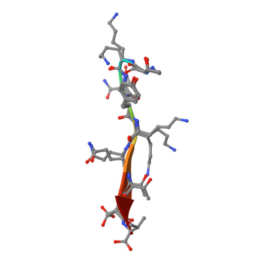
Cysteine-rich PDZ-binding protein9Rattus norvegicusMutation(s): 0 
UniProt
Find proteins for Q792Q4 (Rattus norvegicus)
Explore Q792Q4 
Go to UniProtKB:  Q792Q4
Entity Groups
UniProt GroupQ792Q4
Sequence Annotations
Expand
Reference Sequence

Experimental Data & Validation

Experimental Data

  • Method: X-RAY DIFFRACTION
  • Resolution: 1.35 Å
  • R-Value Free:  0.147 (Depositor), 0.149 (DCC) 
  • R-Value Work:  0.122 (Depositor), 0.122 (DCC) 
  • R-Value Observed: 0.124 (Depositor) 
Space Group: P 41 3 2
Unit Cell:
Length ( Å )Angle ( ˚ )
a = 90.733α = 90
b = 90.733β = 90
c = 90.733γ = 90
Software Package:
Software NamePurpose
PHENIXrefinement
SCALEPACKdata scaling
DENZOdata reduction
PHENIXphasing
PDB_EXTRACTdata extraction

Structure Validation

View Full Validation Report


Entry History 

& Funding Information

Deposition Data


Funding OrganizationLocationGrant Number
National Science Foundation (NSF, United States)United StatesDGE2140743
National Institutes of Health/National Institute of General Medical Sciences (NIH/NIGMS)United StatesDP2-GM141000

Revision History  (Full details and data files)

  • Version 1.0: 2026-11-04
    Type: Initial release