 Download Mendeley
A trefoil knot mimic and multiple pore states define the mechanism of a split-shell Tc toxin
Ng'ang'a, P.N., Raunser, S., Bader, R., Sitsel, O.(2026) Pnas
Experimental Data Snapshot
wwPDB Validation 3D Report Full Report

(2026) Pnas
Entity ID: 1 | |||||
|---|---|---|---|---|---|
| Molecule | Chains | Sequence Length | Organism | Details | Image |
| Virulence plasmid A protein | A [auth D], B [auth E], C, D [auth A], E [auth B] | 2,535 | Photorhabdus luminescens | Mutation(s): 0 Gene Names: SAMN02982990_03606 | 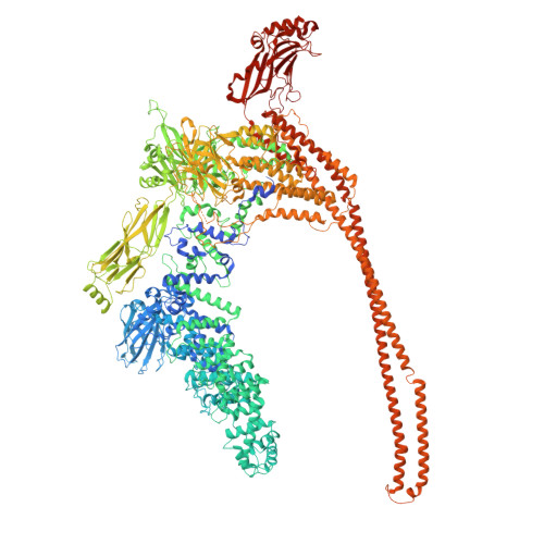 |
UniProt | |||||
Find proteins for A0A1G5RA58 (Photorhabdus luminescens) Explore A0A1G5RA58 Go to UniProtKB: A0A1G5RA58 | |||||
Entity Groups | |||||
| Sequence Clusters | 30% Identity50% Identity70% Identity90% Identity95% Identity100% Identity | ||||
| UniProt Group | A0A1G5RA58 | ||||
Sequence AnnotationsExpand | |||||
Reference Sequence | |||||
| Task | Software Package | Version |
|---|---|---|
| RECONSTRUCTION | cryoSPARC |
| Funding Organization | Location | Grant Number |
|---|---|---|
| Max Planck Society | Germany | -- |
