30AC | pdb_000030ac

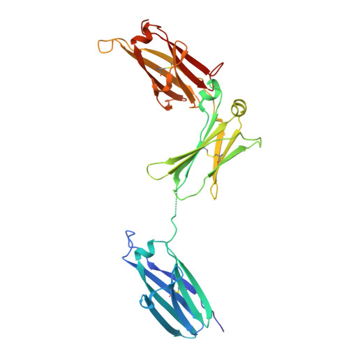
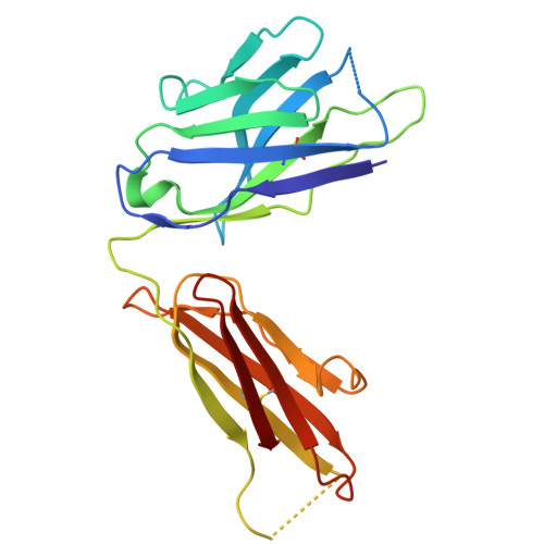
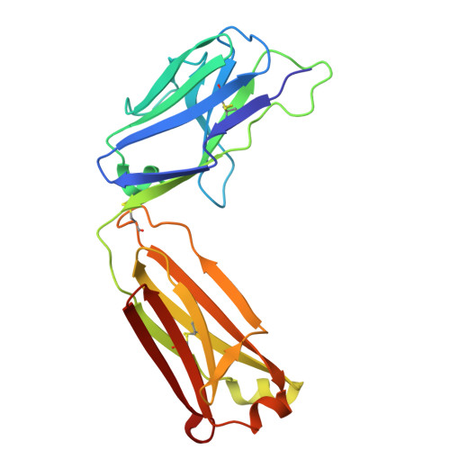
Complex between IgE-Fc and anti-IgE Fab aeFab67


Experimental Data Snapshot

  • Method: X-RAY DIFFRACTION
  • Resolution: 3.20 Å
  • R-Value Free: 
    0.258 (Depositor), 0.258 (DCC) 
  • R-Value Work: 
    0.204 (Depositor), 0.203 (DCC) 
  • R-Value Observed: 
    0.206 (Depositor) 

Starting Models: experimental
View more details

wwPDB Validation 3D Report Full Report

Validation slider image for 30AC

This is version 1.1 of the entry. See complete history

Literature

Antibodies targeting a shared epitope exploit IgE allostery to drive distinct functional outcomes.

Davies, A.M.Drinkwater, N.Beavil, A.J.O'Dowd, V.Lightwood, D.Ceska, T.Henry, A.J.Sutton, B.J.McDonnell, J.M.

(2026) Proc Natl Acad Sci U S A 123: e2613091123-e2613091123

  • DOI: https://doi.org/10.1073/pnas.2613091123
  • Primary Citation Related Structures: 
    30AC, 30AD, 30AE, 30AF

  • PubMed Abstract: 

    The Fc region of immunoglobulin E (IgE-Fc) is an important therapeutic target due to the antibody's critical role in allergic disorders through interactions with high-affinity (FcεRI) and low-affinity (CD23) receptors. IgE-Fc is known to be conformationally flexible, undergoing large-scale structural changes that modulate receptor binding and downstream biological functions. The mechanistic basis for how ligands induce these allosteric changes is not fully understood. In this study, we investigate structure-function relationships of IgE by characterizing four closely related anti-IgE-Fc Fab fragments with highly conserved sequences that recognize a shared epitope at the base of the Cε2 domain. Despite their sequence similarity, these Fabs exhibit markedly different binding characteristics, stoichiometries, and functional activities. Furthermore, the crystal structures of these four Fab complexes reveal that the bound IgE-Fc adopts a wide range of conformations. These structural differences directly dictate the functional outcomes, ranging from conformations that facilitate FcεRI binding to those that prevent or destabilize receptor interaction. Our findings demonstrate that highly similar antibodies binding to the same epitope on a protein that exhibits significant conformational plasticity can elicit distinct and divergent functional outcomes. This work challenges the conventional antibody discovery paradigm, i.e., "same epitope, same functional outcome," and highlights the importance of accounting for both the target's structural flexibility and the resulting functional diversity within antibody selection strategies. This is especially critical when targeting dynamic proteins where allosteric mechanisms are fundamental to function.


  • Organizational Affiliation
    • Randall Centre for Cell and Molecular Biophysics, King's College London, New Hunt's House, London SE1 1UL, United Kingdom.

Macromolecule Content 

  • Total Structure Weight: 164.82 kDa 
  • Atom Count: 11,153 
  • Modeled Residue Count: 1,468 
  • Deposited Residue Count: 1,504 
  • Unique protein chains: 3

Macromolecules

Find similar proteins by:|  3D Structure
Entity ID: 1
MoleculeChains  Sequence LengthOrganismDetailsImage
Isoform 1 of Immunoglobulin heavy constant epsilon
A, B
327Homo sapiensMutation(s): 3 
Gene Names: IGHE
UniProt & NIH Common Fund Data Resources
Find proteins for P01854 (Homo sapiens)
Explore P01854 
Go to UniProtKB:  P01854
PHAROS:  P01854
GTEx:  ENSG00000211891 
Entity Groups
Sequence Clusters30% Identity50% Identity70% Identity90% Identity95% Identity100% Identity
UniProt GroupP01854
Glycosylation
Glycosylation Sites: 1Go to GlyGen: P01854-2
Sequence Annotations
Expand
Reference Sequence
Find similar proteins by:|  3D Structure
Entity ID: 2
MoleculeChains  Sequence LengthOrganismDetailsImage
aeFab67 heavy chainC [auth H],
E [auth C]
211Oryctolagus cuniculusMutation(s): 0 
Entity Groups
Sequence Clusters30% Identity50% Identity70% Identity90% Identity95% Identity100% Identity
Sequence Annotations
Expand
Reference Sequence
Find similar proteins by:|  3D Structure
Entity ID: 3
MoleculeChains  Sequence LengthOrganismDetailsImage
aeFab67 light chainD [auth L],
F [auth D]
214Oryctolagus cuniculusMutation(s): 0 
Entity Groups
Sequence Clusters30% Identity50% Identity70% Identity90% Identity95% Identity100% Identity
Sequence Annotations
Expand
Reference Sequence

Oligosaccharides

Help  
Entity ID: 4
MoleculeChains Length2D Diagram GlycosylationD Interactions
alpha-D-mannopyranose-(1-3)-[alpha-D-mannopyranose-(1-6)]beta-D-mannopyranose-(1-4)-2-acetamido-2-deoxy-beta-D-glucopyranose-(1-4)-2-acetamido-2-deoxy-beta-D-glucopyranoseG [auth E],
H [auth F]
5N-Glycosylation
Glycosylation Resources
GlyTouCan: G22768VO
GlyCosmos: G22768VO
GlyGen: G22768VO

Experimental Data & Validation

Experimental Data

  • Method: X-RAY DIFFRACTION
  • Resolution: 3.20 Å
  • R-Value Free:  0.258 (Depositor), 0.258 (DCC) 
  • R-Value Work:  0.204 (Depositor), 0.203 (DCC) 
  • R-Value Observed: 0.206 (Depositor) 
Space Group: P 21 21 21
Unit Cell:
Length ( Å )Angle ( ˚ )
a = 83.332α = 90
b = 117.542β = 90
c = 191.979γ = 90
Software Package:
Software NamePurpose
PHENIXrefinement
PHENIXrefinement
Aimlessdata scaling
PDB_EXTRACTdata extraction
xia2data reduction
PHASERphasing

Structure Validation

View Full Validation Report


Entry History 

& Funding Information

Deposition Data


Funding OrganizationLocationGrant Number
Medical Research Council (MRC, United Kingdom)United KingdomG1100090

Revision History  (Full details and data files)

  • Version 1.0: 2026-09-02
    Type: Initial release
  • Version 1.1: 2026-09-16
    Changes: Database references