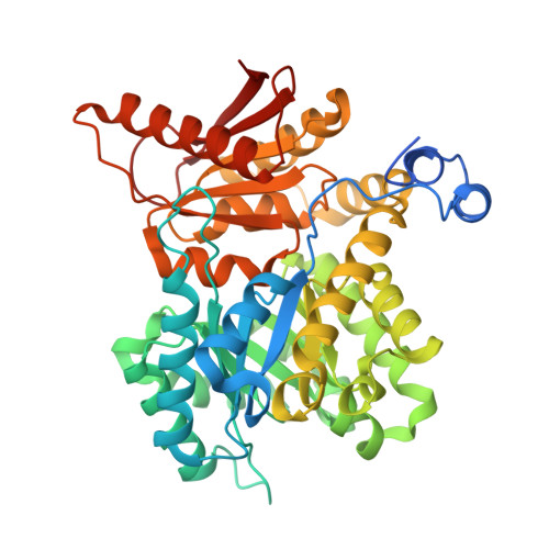
14CQ | pdb_000014cq

Structure of liver pyruvate kinase in complex with allosteric activator 8c


Experimental Data Snapshot

  • Method: X-RAY DIFFRACTION
  • Resolution: 2.18 Å
  • R-Value Free: 
    0.238 (Depositor), 0.225 (DCC) 
  • R-Value Work: 
    0.207 (Depositor), 0.195 (DCC) 
  • R-Value Observed: 
    0.209 (Depositor) 

wwPDB Validation 3D Report Full Report

Validation slider image for 14CQ

Ligand Structure Quality Assessment 


This is version 2.0 of the entry. See complete history. 

Literature

Symmetry-guided fusion converts weak liver pyruvate kinase inhibitors into potent allosteric activators.

Monjas, L., Rodriguez Rivero, A., Fuchtbauer, A.F., Nilsson, O., Haversen, L., Lulla, A., Brear, P., Rutberg, M., Bergh, P.O., Boren, J., Mardinoglu, A., Hyvonen, M., Grotli, M.

(2026) RSC Med Chem 

  • DOI: https://doi.org/10.1039/d6md00604c
  • Primary Citation Related Structures: 
    14CK, 14CL, 14CM, 14CN, 14CO, 14CP, 14CQ, 14CR, 14CS, 14CT, 14CU, 14CV

  • PubMed Abstract: 

    Structure-guided exploitation of symmetry within multimeric protein interfaces offers an opportunity to convert partial ligand-binding poses into higher-affinity modulators. Here, we report a class of symmetrical "Z-shaped" allosteric activators of human liver pyruvate kinase (PKL), generated by symmetry-guided fusion of binding poses identified in inhibitor-bound crystal structures. The resulting ligands span the inter-subunit pocket at the tetrameric dimer-dimer interface and display substantially enhanced activity, with several analogues showing nanomolar activation potency. In HepG2 PKM2-knockout cells, the compounds increased PKL activity, maximal mitochondrial respiratory capacity and glycolytic proton efflux without altering PKL expression, Ser113 phosphorylation-dependent localisation, cell proliferation or triglyceride accumulation under the conditions examined. These findings establish symmetry-guided ligand fusion as a productive design strategy in this PKL system and provide structurally defined chemical tools for investigating the metabolic consequences of direct PKL activation.


  • Organizational Affiliation: 
    • Department of Chemistry and Molecular Biology, University of Gothenburg Medicinaregatan 7b SE-413 90 Gothenburg Sweden grotli@chem.gu.se.

Macromolecule Content 

  • Total Structure Weight: 392.11 kDa 
  • Atom Count: 28,073 
  • Modeled Residue Count: 3,410 
  • Deposited Residue Count: 3,576 
  • Unique protein chains: 1

Macromolecules

Find similar proteins by:|  3D Structure
Entity ID: 1
MoleculeChains  Sequence LengthOrganismDetailsImage
Isoform L-type of Pyruvate kinase PKLR
A, B, C, D, E
A, B, C, D, E, F, G, H
447Homo sapiensMutation(s): 0 
Gene Names: PKLR, PK1, PKL
EC: 2.7.1.40
UniProt & NIH Common Fund Data Resources
Find proteins for P30613 (Homo sapiens)
Explore P30613 
Go to UniProtKB:  P30613
PHAROS:  P30613
GTEx:  ENSG00000143627 
Entity Groups
Sequence Clusters30% Identity50% Identity70% Identity90% Identity95% Identity100% Identity
UniProt GroupP30613
Sequence Annotations
Expand
Reference Sequence

Small Molecules

Ligands 5 Unique
IDChains Name / Formula / InChI Key2D Diagram3D Interactions
WVT
(Subject of Investigation/LOI)

Query on WVT


Download:Ideal Coordinates CCD File
CA [auth E],
HA [auth F],
S [auth C],
X [auth D]
5,5'-(naphthalene-2,6-diyldisulfonyl)di(benzene-1,3-diol)
C22 H16 O8 S2
RKYUAFICCFJCPS-UHFFFAOYSA-N
FBP

Query on FBP


Download:Ideal Coordinates CCD File
AA [auth E]
FA [auth F]
I [auth A]
KA [auth G]
M [auth B]
AA [auth E],
FA [auth F],
I [auth A],
KA [auth G],
M [auth B],
OA [auth H],
Q [auth C],
V [auth D]
1,6-di-O-phosphono-beta-D-fructofuranose
C6 H14 O12 P2
RNBGYGVWRKECFJ-ARQDHWQXSA-N
OXL

Query on OXL


Download:Ideal Coordinates CCD File
BA [auth E]
GA [auth F]
J [auth A]
LA [auth G]
N [auth B]
BA [auth E],
GA [auth F],
J [auth A],
LA [auth G],
N [auth B],
PA [auth H],
R [auth C],
W [auth D]
OXALATE ION
C2 O4
MUBZPKHOEPUJKR-UHFFFAOYSA-L
K

Query on K


Download:Ideal Coordinates CCD File
EA [auth E]
JA [auth F]
L [auth A]
NA [auth G]
P [auth B]
EA [auth E],
JA [auth F],
L [auth A],
NA [auth G],
P [auth B],
RA [auth H],
U [auth C],
Z [auth D]
POTASSIUM ION
K
NPYPAHLBTDXSSS-UHFFFAOYSA-N
MG

Query on MG


Download:Ideal Coordinates CCD File
DA [auth E]
IA [auth F]
K [auth A]
MA [auth G]
O [auth B]
DA [auth E],
IA [auth F],
K [auth A],
MA [auth G],
O [auth B],
QA [auth H],
T [auth C],
Y [auth D]
MAGNESIUM ION
Mg
JLVVSXFLKOJNIY-UHFFFAOYSA-N

Experimental Data & Validation

Experimental Data

  • Method: X-RAY DIFFRACTION
  • Resolution: 2.18 Å
  • R-Value Free:  0.238 (Depositor), 0.225 (DCC) 
  • R-Value Work:  0.207 (Depositor), 0.195 (DCC) 
  • R-Value Observed: 0.209 (Depositor) 
Space Group: C 1 2 1
Unit Cell:
Length ( Å )Angle ( ˚ )
a = 206.922α = 90
b = 112.689β = 92.39
c = 187.888γ = 90
Software Package:
Software NamePurpose
BUSTERrefinement
autoPROCdata processing
XDSdata reduction
Aimlessdata scaling
STARANISOdata scaling
PHASERphasing
PDB_EXTRACTdata extraction

Structure Validation

View Full Validation Report


Ligand Structure Quality Assessment 


Entry History 

Deposition Data

Revision History  (Full details and data files)

  • Version 1.0: 2026-10-14
    Type: Initial release
  • Version 2.0: 2026-11-04
    Changes: Advisory, Atomic model, Data collection, Database references, Derived calculations, Structure summary