Download Mendeley
Structure Basis for Chloride Recognition and Transport by Human Kidney Brain Anion Transporter (KBAT).
Wang, Y., Xie, T., Wu, S., Wang, A., Cheng, W., Huang, J., Yan, R., Chi, X.(2026) Biomolecules 16
- PubMed: 42939639 Search on PubMedSearch on PubMed Central
- DOI: https://doi.org/10.3390/biom16101384
- Primary Citation Related Structures:
9XQF, 9XQU, 9XR3, 9XR4, 9XR5 - PubMed Abstract:
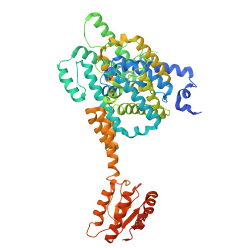
SLC26A11, also known as the kidney and brain anion transporter (KBAT), mediates the transport of multiple anionic substrates across the plasma membrane. It is highly enriched in cerebellar Purkinje cells, where loss of KBAT results in hyperpolarized membrane potentials and increased intrinsic firing frequency, correlating with locomotor dysfunction. Despite its physiological significance, the molecular basis of substrate recognition and transport by KBAT remains elusive. Here, we present the structure of full-length human KBAT in an inward-facing conformation with chloride ions (Cl - ) bound. Sequence analysis of the substrate-binding pockets uncovers the important residue E320 in KBAT. Protonation of E320 abolishes its interaction with S363 and correlates with Cl - binding in the canonical substrate pocket, as substantiated by molecular dynamics (MD) simulations. Quantitative MD contact analysis delineates the rearrangements of residue interactions underlying the alternative access transport, consistent with an elevator-like transport mechanism. The cross-arranged transmembrane helices of the Gate and Core domains require minimal conformational rearrangement to achieve alternating access to the membrane. Collectively, our study provides a critical foundation for comprehensively understanding the recognition and transport mechanisms of human KBAT, with potential implications for developing therapies for neuronal swelling.
- State Key Laboratory of Cellular Stress Biology, School of Life Sciences, Faculty of Medicine and Life Sciences, Xiamen University, Xiamen 361102, China.
Organizational Affiliation: