


Crystal structure of Renilla luciferase variant RLuc8 complexed with Renilla GFP (macromolecular BRET complex)
Marek, M.To be published.
Experimental Data Snapshot
Entity ID: 1 | |||||
|---|---|---|---|---|---|
| Molecule | Chains | Sequence Length | Organism | Details | Image |
| Green fluorescent protein | 231 | Renilla reniformis | Mutation(s): 1 Gene Names: GFP |  | |
UniProt | |||||
Find proteins for Q963I9 (Renilla reniformis) Explore Q963I9 Go to UniProtKB: Q963I9 | |||||
Entity Groups | |||||
| Sequence Clusters | 30% Identity50% Identity70% Identity90% Identity95% Identity100% Identity | ||||
| UniProt Group | Q963I9 | ||||
Sequence AnnotationsExpand | |||||
| |||||
Entity ID: 2 | |||||
|---|---|---|---|---|---|
| Molecule | Chains | Sequence Length | Organism | Details | Image |
| Coelenterazine h 2-monooxygenase | 319 | Renilla reniformis | Mutation(s): 8 EC: 1.13.12.5 | 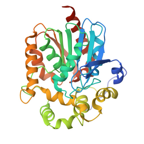 | |
UniProt | |||||
Find proteins for P27652 (Renilla reniformis) Explore P27652 Go to UniProtKB: P27652 | |||||
Entity Groups | |||||
| Sequence Clusters | 30% Identity50% Identity70% Identity90% Identity95% Identity100% Identity | ||||
| UniProt Group | P27652 | ||||
Sequence AnnotationsExpand | |||||
| |||||
| Ligands 1 Unique | |||||
|---|---|---|---|---|---|
| ID | Chains | Name / Formula / InChI Key | 2D Diagram | 3D Interactions | |
| CEI (Subject of Investigation/LOI) Query on CEI | M [auth L] | N-[3-BENZYL-5-(4-HYDROXYPHENYL)PYRAZIN-2-YL]-2-(4-HYDROXYPHENYL)ACETAMIDE C25 H21 N3 O3 CJIIERPDFZUYPI-UHFFFAOYSA-N |  | ||
| Modified Residues 1 Unique | |||||
|---|---|---|---|---|---|
| ID | Chains | Type | Formula | 2D Diagram | Parent |
| CRO Query on CRO | A, B, E, F, I A, B, E, F, I, J | L-PEPTIDE LINKING | C15 H17 N3 O5 |  | THR, TYR, GLY |
| Length ( Å ) | Angle ( ˚ ) |
|---|---|
| a = 96.875 | α = 90 |
| b = 96.875 | β = 90 |
| c = 361.461 | γ = 120 |
| Software Name | Purpose |
|---|---|
| PHENIX | refinement |
| SCALA | data scaling |
| XDS | data reduction |
| PHASER | phasing |
| Funding Organization | Location | Grant Number |
|---|---|---|
| Czech Science Foundation | Czech Republic | GA22-09853S |


