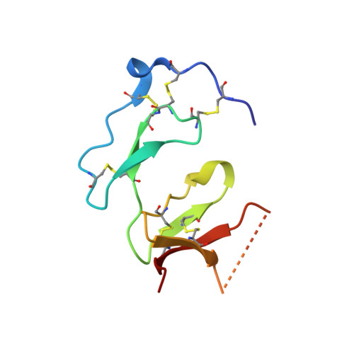
3ZXC | pdb_00003zxc

E69 deletion mutant single insulin-like growth factor binding domain protein (SIBD-1) from Cupiennius salei


Experimental Data Snapshot

  • Method: X-RAY DIFFRACTION
  • Resolution: 1.40 Å
  • R-Value Free: 
    0.191 (Depositor), 0.195 (DCC) 
  • R-Value Work: 
    0.159 (Depositor), 0.159 (DCC) 
  • R-Value Observed: 
    0.161 (Depositor) 

wwPDB Validation   3D Report Full Report


This is version 2.0 of the entry. See complete history


Literature

Structural and Biochemical Characterization of Native and Recombinant Single Insulin-Like Growth Factor-Binding Domain Protein (Sibd-1) from the Central American Hunting Spider Cupiennius Salei (Ctenidae).

Trachsel, C.Widmer, C.Kampfer, U.Buhr, C.Baumann, T.Kuhn-Nentwig, L.Schurch, S.Schaller, J.Baumann, U.

(2012) Proteins 80: 2323

  • DOI: https://doi.org/10.1002/prot.24119

  • PubMed Abstract: 

    Cupiennius salei single insulin-like growth factor binding domain protein (SIBD-1) is an 8.6 kDa Cys-, Pro-, and Gly-rich protein, discovered in the hemocytes of the Central American hunting spider Cupiennius salei. SIBD-1 exhibits high sequence similarity to the N-terminal domain of the insulin-like growth factor-binding protein superfamily and has been reported to play an important role in the spider's immune system. Here, the recombinant expression and the elucidation of the three-dimensional structure of recombinant SIBD-1 and the characterization of the sugar moiety at Thr2 of native SIBD-1 is described in detail.


  • Organizational Affiliation
    • Department of Chemistry and Biochemistry, University of Bern, Bern, Switzerland.

Macromolecules
Find similar proteins by:  (by identity cutoff)  |  3D Structure
Entity ID: 1
MoleculeChains Sequence LengthOrganismDetailsImage
SINGLE INSULIN-LIKE GROWTH FACTOR-BINDING DOMAIN PROTEIN-1
A, B
77Cupiennius saleiMutation(s): 0 
UniProt
Find proteins for G4V4F9 (Cupiennius salei)
Explore G4V4F9 
Go to UniProtKB:  G4V4F9
Entity Groups  
Sequence Clusters30% Identity50% Identity70% Identity90% Identity95% Identity100% Identity
UniProt GroupG4V4F9
Sequence Annotations
Expand
  • Reference Sequence
Experimental Data & Validation

Experimental Data

  • Method: X-RAY DIFFRACTION
  • Resolution: 1.40 Å
  • R-Value Free:  0.191 (Depositor), 0.195 (DCC) 
  • R-Value Work:  0.159 (Depositor), 0.159 (DCC) 
  • R-Value Observed: 0.161 (Depositor) 
Space Group: P 31 2 1
Unit Cell:
Length ( Å )Angle ( ˚ )
a = 49.46α = 90
b = 49.46β = 90
c = 121.499γ = 120
Software Package:
Software NamePurpose
PHENIXrefinement
XDSdata reduction
XSCALEdata scaling
SHELXCDphasing
SHELXEphasing

Structure Validation

View Full Validation Report


Entry History 

Deposition Data

Revision History  (Full details and data files)

  • Version 1.0: 2012-06-06
    Type: Initial release
  • Version 1.1: 2012-08-08
    Changes: Database references
  • Version 2.0: 2024-11-20
    Changes: Atomic model, Data collection, Database references, Derived calculations, Other, Structure summary